The pancreas is an essential organ located behind the stomach in the upper left abdomen. It’s surrounded by other organs like the liver, small intestine, and spleen. Within the body, it plays two roles. The first role is referred to as an endocrine role, where the pancreas secretes hormones that are released into the bloodstream in order to control blood sugar levels. The most commonly known hormones that are released are insulin and glucagon, which lower and raise blood sugar respectfully. The influence of blood sugar regulation can be observed affecting the function of the brain, liver, and kidneys; emphasizing the importance of the pancreas1. The endocrine role takes up about 99% of pancreas function, while the second role, referred to as exocrine function, takes up about 1% of the pancreas’ function2. Exocrine function is crucial to digestion; it produces enzymes that help aid to digestion. Pancreatic juices that contain a concoction of exocrine enzymes are released into the small intestine, where it mixes with stomach acid to help speed up digestion1. While the pancreas is the main location of neuroendocrine tumors, they can also be found in the stomach, specifically in the pylorus. The pylorus is the gateway for food between the stomach and small intestine. The pylorus regulates how much food, in the form of chyme, is allowed to enter the duodenum at one time3.
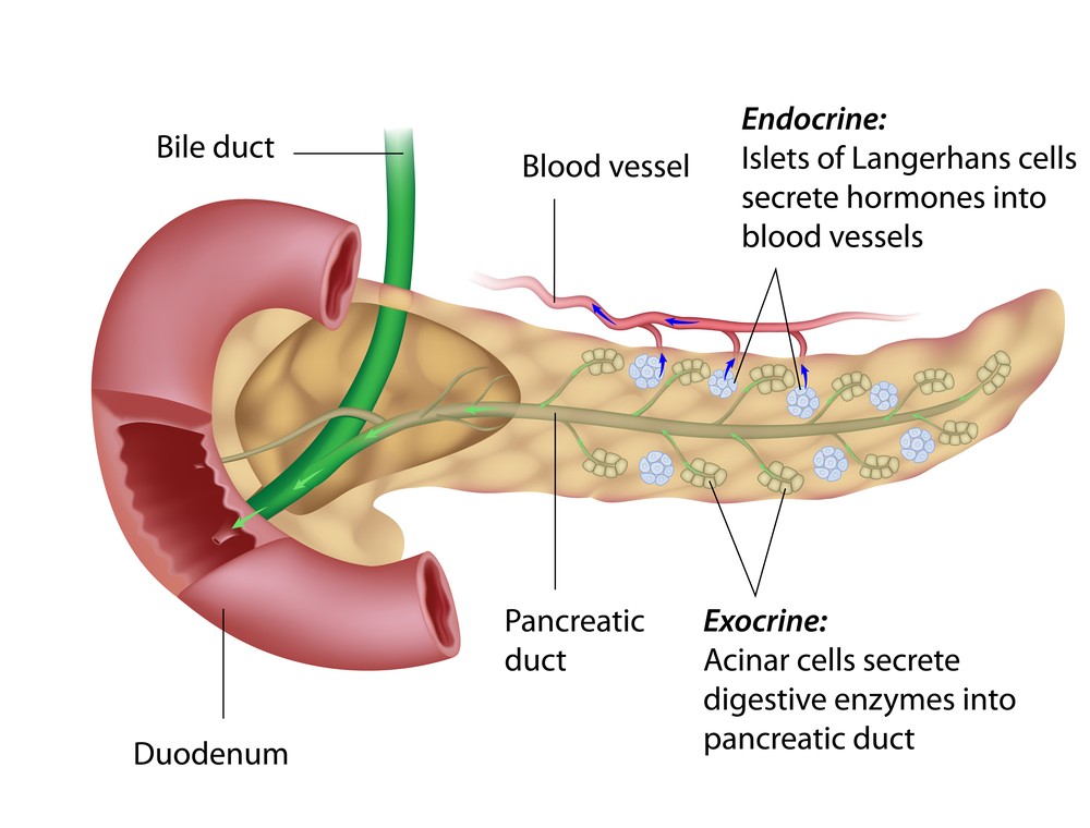
The pancreas has an array of islet cells that produce its own specific protein with its own function. Unless stated otherwise, the location of the cell is the pancreas. The first two cells are the most commonly known pancreatic cells; the ß-cells and α-cells. These two cells produce insulin and glucagon which, as previously mentioned, lower and raise blood glucose levels, respectively4. These two cells work in tandem; when blood glucose levels are high, remaining levels of insulin help to inhibit α-cells and stop the production of glucagon while glucagon does the same thing to ß-cells when blood glucose levels are low. The next cell, the G-cell, is found in the pylorus and produces the hormone gastrin. Gastrin promotes the release of hydrochloric acid (HCl) in order to help create stomach acid4. The last Greek letter cell is the δ-cell. This cell creates somatostatin, which is a secondary inhibitor of endocrine secretions4. Somatostatin serves as an extra regulatory step that works to help promote the release of insulin within ß-cells. When insulin levels are low, somatostatin is released by another intra-pancreatic hormone that binds to δ-cells, resulting in the release of somatostatin. Once somatostatin is released, it binds to ß-cells. This promotes the release of insulin and helps to lower blood sugar5. Non ß-cells are general islet cells of the pancreas that also produce hormonal peptides.
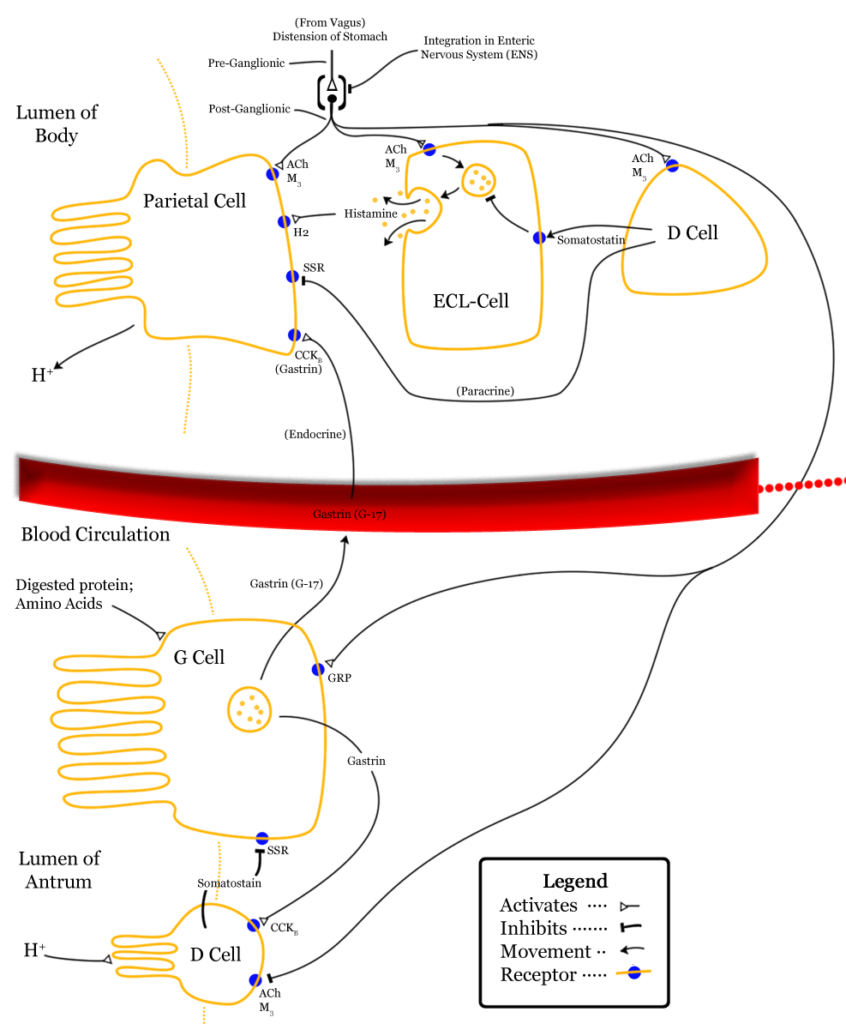
The differentiating factor of islet cells is that they don’t produce specific proteins like all the previously mentioned cells. Non ß-cells have been reported to release vasoactive intestinal peptides (VIP), which help regulate blood flow in the gut4. Islet cells also simulate the release of growth hormone releasing factor (GRF) which help to stimulate the release of growth hormone, as the name suggests4. All these cells help to work together and push forward the function of digestion as well as other regulatory functions throughout the brain, liver, and rest of the body. Of course, these are not all the hormones produced by the pancreas or all the cells that make up the pancreas, however, these are the most researched and best understood in terms of neuroendocrine tumors. So in the best interest of the space, these cells will be the primary focus of this project.
In terms of healthy state pathways and genes, the most understood in pancreatic neuroendocrine tumors focus on death state and cellular growth regulation pathways and genes. This is expected as the actual cells that become tumors continue their programmed function, albeit at a heightened level. The first pathway to address is the apoptotic pathway regulated by the Fas gene. This pathway is regulated by the gene DAXX. DAXX codes for death domain-associated protein 6, which is a protein that acts a histone chaperone and interacts with Fas and centromere protein C6. This interaction helps to push cell death. Later, in the ”A Sign of Disease” section, we will explore the role DAXX plays in the expression of neuroendocrine tumors. Cell death is an important pathway to keep healthy as cancers are characterized by immortal cells that don’t stop proliferation. So, in order to keep healthy pancreatic function, cells must undergo apoptosis when appropriate.
Another important regulatory pathway that helps with general cell regulation that has also been connected to pancreatic neuroendocrine tumor is the mTOR pathway. The mTOR pathway has been shown to be involved in a number of tumors, and recently also shown to be involved in neuroendocrine tumor formation in pre and post clinical trials7. The mTOR pathway has been shown to incorporate serine/threonine kinase that regulates cell growth, cell proliferation, cell motility, cell survival, protein synthesis, autophagy, and transcription. Proliferation is an important regulatory step of cell growth; it bypasses traditional cellular functions in favor of generating cell building blocks to continue rapid growth. This is called the Warburg Effect and is carried out by cancer cells9. Regulation of proliferation is also key to keeping the pancreas in a healthy, functioning state.
Hopefully now you understand the basic functions of a healthy pancreas. If you feel comfortable in your understanding feel free to move forward to the “A Sign of Disease” section in order to understand the ins and outs of pancreatic neuroendocrine tumors!
1 – Wheater’s Functional Histology pp. 287–291
2 – “Pancreas Gland – Endocrine System“. Innerbody. Retrieved 2022-04-15.
3 – Snell, R. S.; Snell, R. S. Clinical Anatomy by Regions, 8th ed.; Wolters Kluwer (Health)/Lippincott Williams & Wilkins: Philadelphia, 2008. 220
4 – StatPearls [Internet]. Treasure Island (FL): StatPearls Publishing; 2022 Jan-. Available from: https://www.ncbi.nlm.nih.gov/pmc/articles/PMC3627221/
5 – DiGruccio, M. R.; Mawla, A. M.; Donaldson, C. J.; Noguchi, G. M.; Vaughan, J.; Cowing-Zitron, C.; van der Meulen, T.; Huising, M. O. Comprehensive Alpha, Beta and Delta Cell Transcriptomes Reveal That Ghrelin Selectively Activates Delta Cells and Promotes Somatostatin Release from Pancreatic Islets. Molecular Metabolism 2016, 5 (7), 449–458. https://doi.org/10.1016/j.molmet.2016.04.007.
6 – Lewis, P. W.; Elsaesser, S. J.; Noh, K.-M.; Stadler, S. C.; Allis, C. D. Daxx Is an H3.3-Specific Histone Chaperone and Cooperates with ATRX in Replication-Independent Chromatin Assembly at Telomeres. Proc. Natl. Acad. Sci. U.S.A. 2010, 107 (32), 14075–14080. https://doi.org/10.1073/pnas.1008850107.
7 – Lamberti, G.; Brighi, N.; Maggio, I.; Manuzzi, L.; Peterle, C.; Ambrosini, V.; Ricci, C.; Casadei, R.; Campana, D. The Role of MTOR in Neuroendocrine Tumors: Future Cornerstone of a Winning Strategy? IJMS 2018, 19 (3), 747. https://doi.org/10.3390/ijms19030747.
8 – Hay, N.; Sonenberg, N. Upstream and Downstream of MTOR. Genes Dev. 2004, 18 (16), 1926–1945. https://doi.org/10.1101/gad.1212704.
9 – Warburg, O. On the Origin of Cancer Cells. Science 1956, 123 (3191), 309–314. https://doi.org/10.1126/science.123.3191.309.
10 – “Beta Cells“. Diabetes Self Management 2016, Retrieved 4.26.2022
11 – “Delta Cells” Wikipedia 2020, Retrieved 4.26.2022
