Cancers and tumors have long been a problem throughout human history. Up until the late 1800s, there was no evidence of what cancer actually was; this was most likely due to its cascade of problems that came along with it. However, starting in 1867, detection and characterization of tumors finally began. Langhans1 in 1867 became the first man to actually describe a tumor. He first noticed lesions on a 50 year old woman who had died of tuberculosis. The lesions were located in the lumen of the small intestine and appeared to be a different type of tissue in comparison to the rest of the intestine. In his 1867 manuscript he focused on the appearance of the tumor he found in the woman, rather than the implications that the different tissue could hold, however, Theodor Langhans had officially laid the groundwork for understanding cancer in humans.
While cancers were being characterized in Germany, around the same time in 1868 the endocrine system was being mapped out. In Prussia during 1868, R. P. Heidenhain founded enterochromaffin (EC) cells, which were cells that he found within the gastric mucosa along with a similar cell he coined as enterochromaffin-like (ECL) cells2; he noted how different the cells were morphologically from other cells found in intestine, specifically their yellow stain from bichromate solutions. The next step in understanding cancer was distinguishing tumor cells from healthy cells. The previous body of work showed that tumor cells display their characteristic under a light microscope and have a tendency to react with silver salts. Jumping from central Europe to western Europe, in 1914 France, Pierre Masson was able to demonstrate that EC and ECL cells were successfully distinguished between tumor cells by absorbing silver staining, showing the argentaffin-staining properties of carcinoid cells3. Masson’s research not only showed that tumors had specific differences that could be detected via cell staining, but also showed that there was a group of cells within the gut that acted differently from lumen cells and served a greater function throughout the body as a whole. Later, in 1928, he determined that the tumor cells he previously stained came from a group of EC and ECL cells that synthesize molecules for neural functions4. This began the understanding of the neuroendocrine system. Later, in 1938, Friederich Feyrter used the same staining techniques that Masson established on healthy cells in the same areas that Masson stained. The healthy in the pancreas cells were unable to absorb the silver staining while tumor cells in the same area were. This difference led Feyrter to conclude that cells Masson stained were pancreatic tumor cells connected to the neuroendocrine system5. Thus, pancreatic neuroendocrine tumors were officially born.
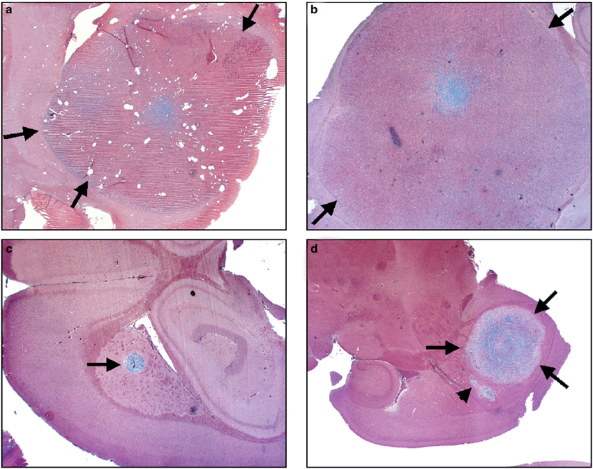
In 1948, 5-hydroxytryptamine (5-HT), or serotonin, was described by M. Rapport4; in 1952, serotonin was isolated through the EC tissues of octopus at the University of Bari, which led to the conclusion that EC cells specifically produced 5-HT. This realization was key to understanding neuroendocrine cells as a whole; by understanding that EC cells were producing one type of signaling molecule, the precedent was established that ECL cells were also synthesizing their own signaling molecules and that there was a better syntax to understanding the EC and ECL cells.
The 1950s saw issues with diagnosing pancreatic neuroendocrine tumors due to their nature. Since the tumors were up regulating metabolic peptides, a cascade of other symptoms would arise with it. This eventually led to a large number of patients being diagnosed as psychotic and eventually dying before the proper diagnosis could be given7. It was not until the 1960s when scientists had finally made clear connections between endocrine cancers and their immediate causes. In 1962 a connection was made between Zollinger-Ellison syndrome and neuroendocrine tumors8. Zollinger-Ellison syndrome is a digestive disorder in which the stomach produces too much gastric acid, which results in abdominal pain, nausea, and other complications. Freisen was able to show that in patients with Zollinger-Ellison syndrome, there existed a tumor, discovered via x-ray, in the EC cell area. This connection helped Freisen establish that Zollinger-Ellison syndrome was being caused by a tumor, specifically tumor cells that up regulate the peptide gastrin, a key peptide that helps promote the release of HCl and create gastric acid for digestion. His conclusion helped to establish gastrinoma as a type of pancreatic neuroendocrine tumor as well as push the classification of pancreatic tumors into a cellular level nomenclature.
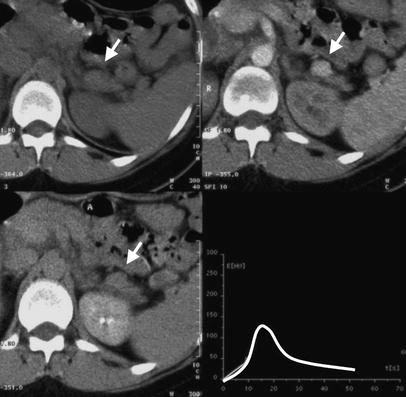
The late 1960s and early 1970s saw a shift in interest from understanding neuroendocrine tumors to actually treating them. One of the most popular drugs to emerge from this era to treat pancreatic tumors was streptozotocin. This particular drug has been shown to be extremely toxic to tumors producing insulin, glucagon, and gastrin in patients that showed symptoms of hyperglycemia9. Streptozotocin would continue to be the leading therapeutic for neuroendocrine tumors, but certain drugs would arise as an attempt to further understand the mechanisms in the pancreas. Combinations of streptozotocin and other compounds like fluorouracil would be given to patients during this time period to view how the combination would affect life span in diagnosed cancer patients. One of the biggest issues with pancreatic neuroendocrine tumors is that being diagnosed is virtually a death sentence; a lot of the drugs from the 1970s aimed to prolong the shortened life spans of its reciprocants as rather than curing them as not enough was known about the cancer itself. The results of streptozotocin along with other therapeutics will be further analyzed within the section “A Variety of Therapeutic Methods.”
The final big advancement in the history of pancreatic neuroendocrine tumors occurred more recently when a mutation at the MEN1 gene, a tumor suppressor in which mutations have been found in 44% of observed tumors10. Understanding that this mutation is present in close to half of all tumors is important in attacking them. In recent years there has been a push to target menin, the protein that MEN1 codes for, in order to inhibit tumor growth. MEN1 and other genes that are involved with pancreatic neuroendocrine tumors will be further explored in the section “A Sign of Disease.” Please enjoy exploring the rest of the website and if you want to move along with the story I’m writing, move next to “A Functioning Pancreas!”
1 – Langhans T: Über einen drüsen polyp im ileum. Virchows Arch Pathol Anat 38:550-560, 1867
2 – Heidenhain R: Untersuchungen über den bau der labdrüsen. Arch f Mikr Anat 6:368, 1870
3 – Gosset, A.; Masson, P. Tumeurs Endocrines de l’appendice. 25:237-240, 1914. Presse Med 1914, 25, 237–240
4 – Modlin, I. M.; Shapiro, M. D.; Kidd, M. Siegfried Oberndorfer: Origins and Perspectives of Carcinoid Tumors. Human Pathology 2004, 35 (12), 1440–1451. Modlin, I. M.; Shapiro, M. D.; Kidd, M. Siegfried Oberndorfer: Origins and Perspectives of Carcinoid Tumors. Human Pathology 2004, 35 (12), 1440–1451. https://doi.org/10.1016/j.humpath.2004.09.018.
5 – Feyrter, F. Über Diffuse Endokrine Epitheliale Organe. Zentralbl Innere Med 1938, 545, 31–41.
6 – Esparmer V, Asero B: Isolation of enteramine from extracts of posterior salivary glands of Octopus vulgaris and of Discoglossus pictus skin. J Biol Chem 200:311-318, 1953
7 – Pearson, O. H.; Pazianos, A. G.; Dominguez, J. M. Neoplastic Disease: Hormone-Producing or Hormone-Dependent Tumors. Annu Rev Med 1960, 11, 243–256. https://doi.org/10.1146/annurev.me.11.020160.001331.
8 – Freisen, S. R.; Tracy, H. J.; Grecory, R. A. Mechanism of the Gastric Hypersecretion in the Zollinger-Ellison Syndrome: Successful Extraction of Gastrin-Like Activity from Metastases and Primary Pancreatico-Duodenal Islet Cell Carcinoma. Annals of Surgery 1962, 155 (2), 167–174 https://doi.org/10.1097/00000658-196200000-00001.
9 – Murraylyon, I. TREATMENT OF MULTIPLE-HORMONE-PRODUCING MALIGNANT ISLET-CELL TUMOR WITH STREPTOZOTOCIN. The Lancet 1968, 292 (7574), 895–898. https://doi.org/10.1016/S0140-6736(68)91058-1.
10 – Jiao, Y.; Shi, C.; Edil, B. H.; de Wilde, R. F.; Klimstra, D. S.; Maitra, A.; Schulick, R. D.; Tang, L. H.; Wolfgang, C. L.; Choti, M. A.; Velculescu, V. E.; Diaz, L. A.; Vogelstein, B.; Kinzler, K. W.; Hruban, R. H.; Papadopoulos, N. DAXX / ATRX , MEN1 , and MTOR Pathway Genes Are Frequently Altered in Pancreatic Neuroendocrine Tumors. Science 2011, 331 (6021), 1199–1203. https://doi.org/10.1126/science.1200609.
11 – Bu, X; Khankaldyyan, V; Gonzales-Gomez, I; Groshen, S; Ye, W; Zhuo, S; Pons, J; Stratton, J; Rosenberg, S; Laug, W; Species-specific Urokinase Receptor Ligands Reduce Glioma Growth and Increase Survival Primarily by an Antiangiogenesis Mechanism. Laboratory investigation; a journal of technical methods and pathology 2004, 85, 667-678.
12 – Pereira, P. L.; Wiskirchen, J. Morphological and Functional Investigations of Neuroendocrine Tumors of the Pancreas. Eur Radiol 2003, 13 (9), 2133–2146. https://doi.org/10.1007/s00330-003-1879-7.

Hi Dave, great post! I thought it was really interesting how the first signaling molecule in the neuroendocrine system to be characterized was serotonin. One cause of major depressive disorder is the poor uptake of serotonin or the lack of serotonin synthesis. During your research, did you come across any publications on the role of serotonin in neuroendocrine cancers and if there is a comorbidity for neuroendocrine cancers and depression?